Integrative Physiology Section
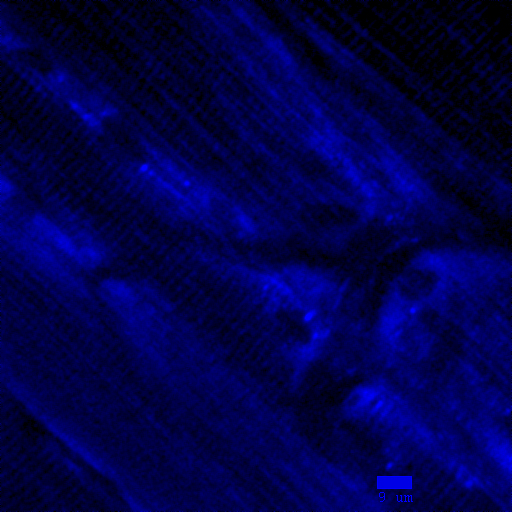
NAD(P)H fluorescence of the tibialis anterior of a living mouse
|
This is an image of NAD(P)H fluorescence of the tibialis anterior of a living mouse taken with two-photon microscopy (pixel size of 0.26 um, excitation 720 nm, emission centered around 450nm). The fluorescence comes from NAD(P)H located in mitochondria which are densely packed in myocytes. The molecule NAD(P)H plays an important role in aerobic mitochondrial respiration and the study of this molecule in living cells gives insight into how aerobic respiration is controlled in muscle. The cross hatch pattern of NAD(P)H fluorescence is from the mitochondria, which lie along the z-lines in skeletal muscle. Both cell nuclei and vessels appear as large non-fluorescent structures in the fluorescence image.
|