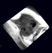
12-5EFICmovie

Episcopic Fluorescence Image Capture
(EFIC)
|
|
|
|
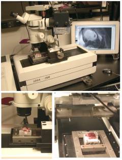
The EFIC system is a setup using a Leica SM2500 microtome,
with a motorized stage, and a Leica MZFLIII microscope mounted on a
customized framework. It serves to
generate snapshots of cross sections of embryos without any shifts in
registration. With these images, full
3D reconstructions can be made to reveal various structural properties of the
heart. The EFIC setup is shown on the
left.
|
Movie Gallery
Cross sectional Movies
Photo Gallery
|
|
|
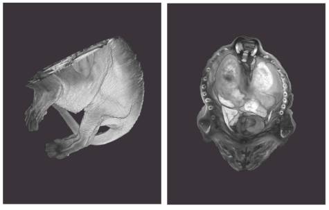
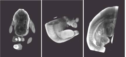
E16.5 Mouse Embryo
3D Reconstructions of a E16.5 day mouse embryo. The images are rotated to different
angles. Full reconstructions were
done on Volocity software (Improvision) from the 3D cross sections generated
by EFIC.
|
|
|
|
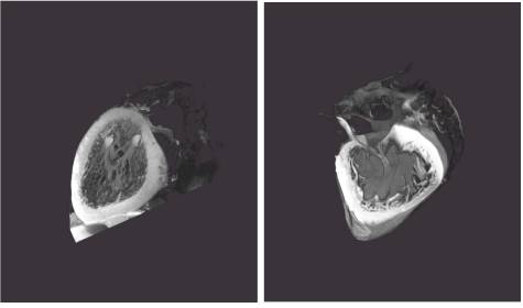
E17.5 Fetal Mouse
Heart
3D reconstructions of a wildtype E17.5 fetal mouse
heart. (Left) Left ventricles showing
2 papillary muscles (Right) Right Ventricle showing the tricuspid valve
|
(A)
(B)
|
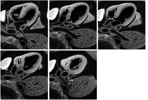
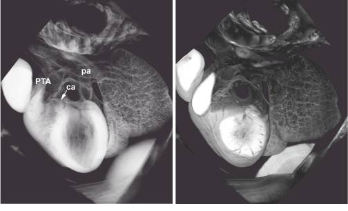
Persistent Truncus Arteriosus (PTA)
These are a series of images, taken from family 53 ENU
mouse neonate, generated in OpenLab (Improvision) using EFIC for phenotyping
purposes. (A) This composite indicates
persistent truncus arteriosus (PTA).
Labels: Ventricular Septal
Defect (VSD), Left Coronary Artery (LCA); Right Coronary Artery (RCA),
Ascending Aorta (AA), Left Pulmonary Artery (LPA); (B) This is the 3D
reconstruction of the cross sections generated. (Left) This shows the diagnosis of PTA off of the right
ventricle. (Right) This shows the
right ventricle and right atria.
|
|
|
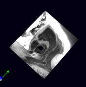
Standard Views for EFIC Reconstructions
This composite demonstrates the capabilities of Mouse
Edinburgh Software where virtual sectioning can be done. It helps in creating reconstructions that
can show structures in different planes.
(Left) Original images sectioned in the Apical view of the E11.5
embryo. (Middle) Virtual sections to
create a Transverse view of the E11.5 embryo. (Right) Virtual
sections to create a Sagittal view of the E11.5.
|
Using Polyethylene Glycol (PEG) as Embedding Medium
E16.5 Fetal Mouse
Heart
3D reconstruction of a E16.5 fetal mouse heart embedded
using PEG.
Home
|
|
 |
Laboratories
of the NHLBI |
|
LDB Home |
 |
|
|
|
|
|
<%
if NIHintranet = "False" and intranet = "Yes" then
'response.write("NIHintranet="+NIHintranet)
' do nothing...
else%>
|
Connexin Regulation of Cell Motility |
|
|
<%
end if
intranet = "No"
%>
<%
if NIHintranet = "False" and intranet = "Yes" then
'response.write("NIHintranet="+NIHintranet)
' do nothing...
else%>
|
|
<%
end if
intranet = "No"
%>
|
|
<%
if NIHintranet = "False" and intranet = "Yes" then
'response.write("NIHintranet="+NIHintranet)
' do nothing...
else%>
|
Connexin Protein Interactions and Cell Signaling |
|
|
<%
end if
intranet = "No"
%>
<%
if NIHintranet = "False" and intranet = "Yes" then
'response.write("NIHintranet="+NIHintranet)
' do nothing...
else%>
|
|
<%
end if
intranet = "No"
%>
|
|
<%
if NIHintranet = "False" and intranet = "Yes" then
'response.write("NIHintranet="+NIHintranet)
' do nothing...
else%>
|
Mouse ENU Mutagenesis |
|
|
<%
end if
intranet = "No"
%>
|
|
<%
if NIHintranet = "False" and intranet = "Yes" then
'response.write("NIHintranet="+NIHintranet)
' do nothing...
else%>
|
Mouse Heart Atlas |
|
|
<%
end if
intranet = "No"
%>
<%
if NIHintranet = "False" and intranet = "Yes" then
'response.write("NIHintranet="+NIHintranet)
' do nothing...
else%>
|
|
<%
end if
intranet = "No"
%>
|
|
<%
if NIHintranet = "False" and intranet = "Yes" then
'response.write("NIHintranet="+NIHintranet)
' do nothing...
else%>
|
Photo/Movie Gallery |
|
|
<%
end if
intranet = "No"
%>
<%
if NIHintranet = "False" and intranet = "Yes" then
'response.write("NIHintranet="+NIHintranet)
' do nothing...
else%>
|
|
<%
end if
intranet = "No"
%>
|
|
<%
if NIHintranet = "False" and intranet = "Yes" then
'response.write("NIHintranet="+NIHintranet)
' do nothing...
else%>
|
Protocols |
|
|
<%
end if
intranet = "No"
%>
|
|
<%
if NIHintranet = "False" and intranet = "Yes" then
'response.write("NIHintranet="+NIHintranet)
' do nothing...
else%>
|
LDB Staff |
|
|
<%
end if
intranet = "No"
%>
<%
if NIHintranet = "False" and intranet = "Yes" then
'response.write("NIHintranet="+NIHintranet)
' do nothing...
else%>
|
|
<%
end if
intranet = "No"
%>
|
|
|
|
|
|
|

